There are several common eye problems including:
Amblyopia (lazy eye). In an infant or a child, the brain will not tolerate double images and will shutdown the vision
in the weaker eye. This involuntary loss of vision is called "lazy eye"
or amblyopia. Here's another way to say it: Amblyopia is a healthy eye
that does not see. Only infants and children develop amblyopia; and the
vision loss can be reversed with therapy if the contributing eye problem
is corrected during childhood.
Amblyopia is a serious problem for your children. So long as the
underlying eye problem remains untreated, the vision in the weaker eye
does not develop fully. Lazy eye can also result from other eye
problems, such as ptosis (drooping of the eyelid) or a significant
refractive error in one eye. If detected early amblyopia can be
corrected with patching and/or eye drops applied to the better eye -
forcing the weaker eye to recover useful function.
Presbyopia.
This is the loss of the ability to see close objects or small print
clearly. It is a normal process that happens slowly over a lifetime, but
you may not notice any change until after age 40. Presbyopia is often
corrected with reading glasses. Bifocal glasses permit the wearer to see
objects clearly, both near and distant.
Floaters.
These are tiny spots or specks that float across the field of vision.
Most people notice them in well-lit rooms or outdoors on a bright day.
Floaters are often considered normal, but can sometimes indicate a more
serious eye problem. These include conditions such as a retinal
detachment, especially if floaters are accompanied by light flashes, or
any reduction in your field of vision, like a curtain falling over the
eye. If you notice a sudden change in the type or number of spots or
flashes you see, seek medical advice as soon as possible
Dry eyes.
This happens when tear glands cannot make enough tears or produce poor
quality tears. Dry eyes can be uncomfortable, causing itching, burning
or, rarely, some loss of vision. Your doctor or eye specialist may
suggest using a humidifier in your home, special eye drops that simulate
real tears, or plugs that are placed in tear ducts to decrease tear
drainage. Surgery may be needed in more serious cases of dry eyes.
Tearing.
Having too many tears can come from being sensitive to light, wind or
temperature changes. Protecting your eyes by shielding them or wearing sunglasses
can sometimes solve the problem. Tearing may also mean that you have a
more serious problem, such as an eye infection or a blocked tear duct.
Your doctor or eye specialist can offer advice about treatment for these
conditions.
Cataracts. Cataracts are cloudy areas that
develop within the eye lens. Since the lens in a healthy eye is clear
like a camera lens, light has no problem passing through the lens to the
back of the eye to the retina where images are processed. When a
cataract is present, the light cannot get through the lens as easily
and, as a result, vision can be impaired. Cataracts often form slowly,
causing no pain,
redness or tearing in the eye. Some stay small and do not alter
eyesight. If they become large or thick and affect vision, cataracts can
usually be treated with surgery to replace the lens.
Colour blindness. Colour
blindness is most commonly a disorder of the retina's light-sensitive
photoreceptor cells, which respond to different coloured light rays.
There are two kinds of photoreceptors: cones work best in bright light
and rods work best in dim light. Each photoreceptor produces pigments
that respond to specific colours of light. Colour vision is affected if
those pigments are absent, defective, or if they respond to the wrong
wavelengths.
You have probably seen how paint colours can be mixed. Colour vision
works much the same way because visible light is a mixture of different
light rays (wavelengths). Colour perception problems occur more often in
men, and affects approximately one in 20 men in the UK. It is much
less common in women, affecting only one in 200 women. It is extremely
rare for someone to be totally colour-blind, that is, able to see only
shades of grey.
Conjunctivitis.
The conjunctiva -- the moist, transparent membrane that covers the
eyeball and your inner eyelid -- can become inflamed for various
reasons. It can cause redness, itching, burning, tearing, discharge or a
feeling of something in the eye. Conjunctivitis occurs in people of all
ages and can be caused by infection, exposure to chemicals and
irritants, or allergies.
Most cases of conjunctivitis run a predictable course, and the
inflammation usually clears up in a few days. Although infectious
conjunctivitis can be highly contagious, it is rarely serious and will
not permanently harm your vision if detected and treated promptly.
Corneal diseases.
The cornea is the clear, dome-shaped "window" at the front of the eye.
It helps to focus light that enters the eye. Disease, infection, injury
and exposure to toxic agents can damage the cornea causing eye redness,
watery eyes, pain, reduced vision or a halo effect. Treatments include
making adjustments to the eyeglass prescription, using medicated eye
drops or having surgery.
Eyelid problems. The eyelids
protect the eye, distribute tears and limit the amount of light entering
the eye. Pain, itching, tearing and sensitivity to light are common
symptoms of eyelid problems. Other problems may include drooping
eyelids, blinking spasms or inflamed outer edges of the eyelids near the
eyelashes. Eyelid problems often can be treated with proper cleaning, medication or surgery.
Eyestrain.
Eyestrain is discomfort due to an uncorrected refractive problem. This
common vision problem may occur while performing distant visual
activities like driving or watching a film or during-close-up tasks.
Familiar symptoms of eyestrain include headache, brow-ache, eye fatigue
and/or a pulling sensation. Eyestrain quickly goes away if the
refractive problem is resolved. Prolonged focusing can lead to
eyestrain, such as working at the computer for hours. Children have a
far more flexible focusing capacity. How often do you ever hear a child
complain of eyestrain while playing video games? If you wear
prescription glasses, recurring eyestrain may be an indication that you
need updated glasses or a new prescription. Eye exercises or resting the
eyes every 30 minutes helps relieve eyestrain, especially when working
with computers.
Glaucoma.
This condition develops when there is too much fluid pressure inside
the eye. Glaucoma occurs when the normal flow of the watery fluid cannot
drain properly. If not treated early, this can lead to permanent vision
loss and blindness. Glaucoma is less commonly caused by other factors
such as injury to the eye, severe eye infection, blockage of blood
vessels or inflammatory disorders of the eye. Because most people with
glaucoma have no early symptoms or pain, it is very important to get
your eyes checked regularly. Treatment may include prescription eye
drops, oral medications or surgery.
Macular degeneration. Because
the symptoms usually do not appear in people under 55 years of age, the
disorder is more accurately referred to as age-related macular
degeneration (AMD). Approximately 2% of people over 50 years of age have
age-related macular degeneration. In people over 65 years of age, the
number rises to 8%, with about 20% of those over 85 years of age having
the condition.
AMD affects your central vision, meaning if you were looking at a
photograph, you would not be able to see the middle of the picture but
could still see the edges (preserved peripheral vision). If you are over
65, macular degeneration may already affect your central vision -- the
vision you need for reading and close work like sewing. The disorder
occurs in two forms, dry and wet. The less common wet form of AMD
requires immediate medical attention. Any delay in treatment may result
in loss of your central vision.
Night blindness. Night
blindness -- difficulty seeing in dim light -- occurs when rod
photoreceptor cells begin to deteriorate. Rods work best in low light.
There are many different forms of night blindness, but it may be linked
to liver disorder, vitamin-A deficiency, inherited disease of the retina, such as retinitis pigmentosa.
Retinal disorders.
The retina is a thin lining on the back of the eye made up of cells
that collect visual images and pass them on to the brain. Retinal
disorders interrupt this transfer of images. They include age-related
macular degeneration, diabetic retinopathy
and retinal detachment. Early diagnosis and treatment of these
conditions is important to maintain vision. Although a detached retina
is not painful, it is a medical emergency. If the retina is not
reattached to the eye wall promptly, retinal cells starve and permanent
blindness can result. Risk factors for retinal detachment include
moderate or extreme short-sightedness, previous eye surgery or injury,
previous retinal detachment and/or Inherited thinness of retinal tissue.
Strabismus (squint).
The medical term for misaligned eyes is strabismus. If strabismus
(squint) develops in an adult, perhaps after a trauma to the head or
after a stroke,
the person is likely to experience double vision. Double vision occurs
because the two eyes are looking at different images. Did you know that
there are six different muscles that are attached to each eye to help it
turn and rotate? The eyes may not appear straight because one or more
muscles are pulling too hard or other muscles are too weak. If the eyes
turn inward leading to "crossed eyes" we call it esotropia. If they turn
outward, called "wall eyes", then the condition is labelled exotropia.
There are different treatments for strabismus depending on the specific
cause. Some cases are managed with eye muscle surgery, some simply need
glasses.
Temporal arteritis. This condition is an inflammation of the arteries in the temple area of the forehead. It can begin with a severe headache, pain when chewing, and tenderness or swelling
in the temple area. It may be followed in a few days or weeks by sudden
vision loss -- usually in one eye. Other symptoms can include shaking,
weight loss
and low-grade temperature. Scientists don't know the cause of temporal
arteritis but they think it may be caused by an impaired immune system.
Sudden vision loss in the other eye may occur within a few days or weeks
of the first eye. Getting to an ophthalmologist
-- an eye specialist -- whenever sudden vision loss occurs is critical.
Early treatment with medication may help prevent vision loss in one or
both eyes.

Friday, 24 January 2014
Thursday, 16 January 2014
Eye Floaters
Eye floaters are small moving spots that appear in your
field of vision. They may be especially noticeable when you look at
something bright, such as white paper or a blue sky.
Eye floaters can be annoying, but they generally don't interfere with your sight.
Occasionally a particularly large eye floater may cast a
subtle shadow over your vision. But this tends to occur only in certain
types of light.
Most of the time people learn to live with eye
floaters and ignore them. And they often improve over months to years.
Only rarely do benign eye floaters become bothersome enough to consider
treatment.
But sometimes eye floaters are a sign of a more
serious condition. You should seek immediate medical attention if you
notice a sudden increase in the number of eye floaters.
Immediate medical attention is especially important
if the floaters are accompanied by flashes of light or a loss of side
vision. If you have these symptoms, see an eye doctor right away.
Without immediate treatment, you can have permanent vision loss. These
symptoms may be caused by:
- Retinal detachment
- Retinal tear
- Bleeding within the eye
Symptoms of Eye Floaters
Eye floaters move as the eyes move. They generally appear to dart away when you try to focus on them.
Eye floaters can appear in many different shapes, such as:
- Black or gray dots
- Squiggly lines
- Threadlike strands, which can be knobby and semi-transparent
- Cobwebs
- Ring shaped
Once you develop eye floaters they usually do not go away, though they tend to improve over time.
Causes of Eye Floaters
Most eye floaters are caused by small flecks of a protein called collagen.
The back compartment of the eye is filled with a gel-like substance called vitreous humor.
As you age, the vitreous and its millions of fine
collagen fibers shrink and become shred-like. Shreds can accumulate in
the vitreous. This can cause a change in the amount of light that hits
the retina -- the light-sensitive tissue in the back of the eye. This
change causes the symptoms of eye floaters.
These changes can happen at any age. They most often
occur between ages 50 and 75, especially in people who are very
nearsighted or have had cataract surgery.
Rarely, eye floaters can result from other eye surgery or:
- Eye disease
- Eye injury
- Diabetic retinopathy
- Crystal-like deposits that form in the vitreous
- Eye tumors such as lymphoma (rarely)
Serious eye disorders associated with eye floaters include:
- Retinal detachment
- Retinal tear
- Vitreous hemorrhage (bleeding)
- Vitreous and retinal inflammation caused by viral infections, fungal infections, or auto-immune inflammation
- Eye tumors
In addition, a unique form of eye floaters is associated with the visual aura of migraine headaches.
When to Seek Medical Attention for Eye Floaters
If you only have a few eye floaters that don't change over time, it usually does not indicate a serious eye problem.
It's important to see a doctor if:
- Eye floaters seem to worsen over time, especially if the changes are sudden in onset.
- You experience flashes of light or any vision loss accompanied by eye floaters.
- You develop eye floaters after eye surgery or eye trauma.
- You have eye pain along with eye floaters.
Monday, 13 January 2014
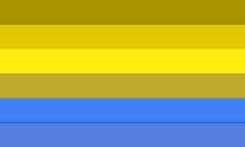
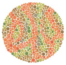
What is Color-Blindness
Color-blindness is the inability to distinguish the differences between certain colors. This condition results from an absence of color-sensitive pigment in the cone cells of the retina, the nerve layer at the back of the eye.
Most color vision problems are inherited and are present at birth. Approximately 1 out of 12 males and 1 out of 20 women are color blind.
What does a color-blind person see?
A
person with color-blindness has trouble seeing red, green, blue, or
mixtures of these colors. The most common type is red-green
color-blindness, where red and green are seen as the same color.
Here are some illustrations of the most common forms of color-blindness:
 |  |
The colors of the rainbow
Normal color vision | The colors of the rainbow Deuteranope (simulation) Absence of green retinal photoreceptors |
 |  |
| The colors of the rainbow Protanope (simulation) Absence of red retinal photoreceptors. | The colors of the rainbow Tritanope (simulation) Absence of blue retinal receptors |
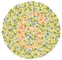
Tests for Color-Blindness
The typical test for color-blindness is based on a person's ability to see numbers inside a circle.Before you look at these tests, be aware of the following:
1.
Your computer and monitor may not display the colors accurately. If you
cannot see the number that does not necessarily mean you are
color-blind. The numbers may more visible with some resolutions and
display settings than with others.
2.
The results of this test are not to be considered a valid medical test
for color blindness and merely serve to illustrate the tests available.
If you have any questions about your own possible color vision
deficiencies consult a licensed medical professional.
Here is a sample of charts used to test for color-blindness.
There is a number in the center of the circle.
If you can see the number, chances are you are not color-blind.
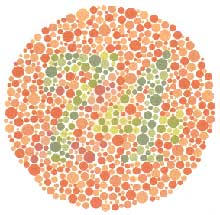 |  |
| Plate 1 What number do you see? | Plate 2 What number do you see? |
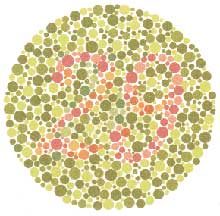 |  |
| Plate 3 What number do you see? | Plate 4 What number do you see? |
Here are the answers:
Plate 1Those with normal color vision should read the number 74. Plate 2 Those with normal color vision should read the number 6. Plate 3 Those with normal color vision should read the number 29. Those with red-green deficiencies read the number 70. Those with total color-blindness can not read any number. Plate 4 Those with normal color vision should not be able to read any number. Most of those with red-green deficiencies should read the number 5. Those with total color -blindness can not read any number. |
Friday, 10 January 2014
Can Sunlight Damage a Toddler's Eyes? by Kay Ireland, Demand Media
You slather your toddler's sensitive skin in sunscreen to protect
her from the sun's rays, but your toddler's eyes can be just as
sensitive to the sun and there's no lotion that can help. By
understanding the ways the sun can damage your toddler's eyes, you can
choose protection to keep his eyes safe and healthy.
Short-term Risks
Children are especially at risk for sun damage to the eyes
because they typically spend more time in the sun, according to the
American Optometric Association. The sun emits UV rays, which are a type
of radiation. When exposed to high amounts of UV rays, a toddler can
experience photokeratitis, which is essentially a sunburn of the eye.
The condition is short-lived and causes mild pain and discomfort, such
as a gritty and burning sensation in the eyes.
Long-term Issues
While photokeratitis doesn't have any lasting effects on your
toddler's eye, excessive and frequent exposure to UV rays can have
long-term effects. The AOA warns that too much sun over time can result
in the possibility of a cataract and could cause damage to the retina,
both which can result in vision problems and the necessity for surgery
in the future. The more your toddler's eyes are exposed to UV rays, the
higher his risk for sun-related vision issues in the future.
Eye Protection
While you can't put sunscreen on a child's eyes, you can use
methods to protect your toddler's vision. Some toddlers are happy to
slip on a pair of UV-protection sunglasses -- look for wraparound models
that protect the entire eye and that stay on your toddler's head
easily. If your little one won't wear sunglasses, try a hat with wide
brim to keep the sun out of his eyes while he's busy playing, suggests
the Skin Cancer Foundation.
Sun Smarts
While you don't need to hide from the sun, it's important that
your toddler learns how to be sun smart from the start. By cautioning
him against looking directly at the sun, finding shade and explaining
the use of sunscreen, sunglasses and hats, your toddler can become more
enthusiastic about sun safety and less likely to damage to his sensitive
eyes. According to the American Academy of Pediatrics, one-quarter of
lifetime sun exposure happens during childhood. By teaching your toddler
sun safety, he can play outside and you can worry less.
Can Wearing Glasses Make a Toddler's Eyesight Better? by Kristen Berry, Demand Media
Wearing glasses can markedly improve a toddler's vision. According
to the American Association for Pediatric Ophthalmology and
Strabismus, the first five to six years of a child's life involves major
vision growth and development. Not only can glasses improve your
toddler's vision, they might play an important role in ensuring normal
development of his vision.
Myopia -- Nearsightedness
According to research at Boston Children's Hospital, if your
toddler has trouble seeing faces from across a room, or character's
faces on a movie theater screen, she might be nearsighted.
Nearsightedness is caused by an eyeball that is too long. This irregular
shape causes incoming light rays to focus in front of your toddler's
retina instead of directly on it. Distant objects appear blurry. If your
toddler is nearsighted, prescription glasses will refocus the incoming
light and create clearer images.
Hyperopia -- Farsightedness
Your toddler will likely have difficulty in expressing her vision
problems orally. Look for signs such as the inability to fixate on or
follow objects. According a nonprofit health group, Fairview Health
Services in Minneapolis, if objects that are close to your toddler's
line of immediate vision cause her to squint, she might be farsighted,
also called hyperopia. Hyperopia is the refractive defect in which a
shorter eyeball causes the image of an object to be focused behind the
retina. Eyeglasses will help to correct or improve your toddler's
hyperopia through adjusting the focusing power to the retina. Your
toddler will have an easier time seeing close objects more clearly.
Astigmatism
Astigmatism is a common condition in which your toddler might
have an abnormal curvature of her cornea. This can cause two focal
points to fall in two different locations, whether your toddler is
seeing an object up close, or at a distance. This can put a strain on
your toddler's eyes and even cause her undue fatigue. According to
Boston Children's Hospital, if your toddler has astigmatism, wearing
glasses will make her vision sharper and more consistent.
Dependency
You might worry that your toddler's need for vision correction
could cause her to become dependent on glasses and worsen her vision.
According to the American Association for Pediatric Ophthalmology and
Strabismus, the opposite might be true. If your toddler does not
consistently wear the glasses prescribed, her normal vision development
can be inhibited or even adversely affected. The American Association
for Pediatric Ophthalmology and Strabismus recommends finding an
optician who is experienced in pediatric eyewear. Your toddler's frames
should fit comfortably with the eye centered in the middle of the lens.
Glasses and Contact Lenses
Why do some people need glasses and others don't?
Everyone's eyes are a little different — not just the color, but the way they work and how well they see. Sometimes all the parts of the eye don't work together the way they should. But eyeglasses or contact lenses, also called corrective lenses, can help most people see more clearly.
When all of the eyes' parts are working properly, a kid doesn't have vision problems. You can see because your eyes capture an image like a camera and send that image to your brain, where it can be interpreted. For instance, if there's an elephant in front of you, almost instantly, your brain says, "Hey, that's an elephant."
Your eyes need to bend light rays so the image can be focused sharply on your retina. The better your retina records the image, the more likely that your brain will interpret the image, and the more likely you will see the image clearly.
Refracting is a big word that means bending light rays. If a person has vision trouble, it's often a refractive problem. Glasses or contact lenses work so well because they can correct refractive problems. In other words, they bend the light rays in a way that lets you see more clearly.
Laser surgery also can correct some vision problems, but it's not recommended for kids because they're still growing.
Another refractive problem is called astigmatism. This means that the cornea is an uneven shape, and it bends the light in different directions. This can distort what a person sees and make things look blurry.
Glasses or contact lenses correct vision because they allow the eye to focus light in the right spot on the retina — the spot that produces the clearest image. Because everyone's eyes are different, a pair of glasses that makes one person see wonderfully may look terribly blurry to someone else. You know this if you've ever tried on somebody else's glasses!
If you need glasses or contact lenses, your doctor will write you a prescription. In this case, a prescription doesn't mean medicine you'll pick up at the drugstore. A vision prescription is a piece of paper with numbers on it. The people who will make your glasses for you need these numbers to create lenses that will correct the way your eye bends light. Remember, the target is right in the center of the retina.
If you're going to get glasses, it's time to pick frames. It can be fun to try these on. Choose ones that are comfortable and sturdy. But also make sure you like them — you'll be wearing them a lot!
The lenses themselves can be made of different materials, such as safety glass or shatterproof plastic (polycarbonate). Because glass tends to be heavy and it may shatter, most glasses today are made of polycarbonate plastic. If you play sports, you may want to ask about special eyewear you can wear on the field.
Glass tends to be heavy and it can shatter. Plastic scratches easily, but it's often the best choice for kids. If you play sports, you may want to ask about eyewear you can wear on the field.
With glasses, you'll also want to find out how to clean them properly. And it helps if you have a glasses case and put them in it when you're not wearing them. The last thing you want is to sit on your new glasses.
If you're going to get contact lenses, you'll get some advice from the ophthalmologist or optician about which kind will be best for you. Some are disposable and others need to be cleaned. When you learn which type you're going to get, you can start becoming an expert in how to wear them safely and keeping them clean. The most important thing about contact lenses is good hygiene to prevent infections in your eye.
But the really fun part of new glasses or contact lenses is how well you can see. They can make your whole world look better!
Everyone's eyes are a little different — not just the color, but the way they work and how well they see. Sometimes all the parts of the eye don't work together the way they should. But eyeglasses or contact lenses, also called corrective lenses, can help most people see more clearly.
How Eyes Work
The eyeball includes the cornea, clear tissue that helps the eye focus; the iris, the colored part; the pupil, which lets light into the eye; the lens, which also helps the eye focus; and the retina, at the very back of the eye.When all of the eyes' parts are working properly, a kid doesn't have vision problems. You can see because your eyes capture an image like a camera and send that image to your brain, where it can be interpreted. For instance, if there's an elephant in front of you, almost instantly, your brain says, "Hey, that's an elephant."
Your eyes need to bend light rays so the image can be focused sharply on your retina. The better your retina records the image, the more likely that your brain will interpret the image, and the more likely you will see the image clearly.
Refracting is a big word that means bending light rays. If a person has vision trouble, it's often a refractive problem. Glasses or contact lenses work so well because they can correct refractive problems. In other words, they bend the light rays in a way that lets you see more clearly.
Laser surgery also can correct some vision problems, but it's not recommended for kids because they're still growing.
Nearsighted and Farsighted — Which Is Which?
Nearsightedness and farsightedness are common refractive problems. It's easy to get the two confused:- Nearsighted means someone can see stuff that's near, like a book, but has trouble seeing stuff that's far away.
- Farsighted means someone can see stuff that's far away, but has trouble seeing up close, like reading the print in a book.
Another refractive problem is called astigmatism. This means that the cornea is an uneven shape, and it bends the light in different directions. This can distort what a person sees and make things look blurry.
Glasses or contact lenses correct vision because they allow the eye to focus light in the right spot on the retina — the spot that produces the clearest image. Because everyone's eyes are different, a pair of glasses that makes one person see wonderfully may look terribly blurry to someone else. You know this if you've ever tried on somebody else's glasses!
If you need glasses or contact lenses, your doctor will write you a prescription. In this case, a prescription doesn't mean medicine you'll pick up at the drugstore. A vision prescription is a piece of paper with numbers on it. The people who will make your glasses for you need these numbers to create lenses that will correct the way your eye bends light. Remember, the target is right in the center of the retina.
If you're going to get glasses, it's time to pick frames. It can be fun to try these on. Choose ones that are comfortable and sturdy. But also make sure you like them — you'll be wearing them a lot!
The lenses themselves can be made of different materials, such as safety glass or shatterproof plastic (polycarbonate). Because glass tends to be heavy and it may shatter, most glasses today are made of polycarbonate plastic. If you play sports, you may want to ask about special eyewear you can wear on the field.
Glass tends to be heavy and it can shatter. Plastic scratches easily, but it's often the best choice for kids. If you play sports, you may want to ask about eyewear you can wear on the field.
With glasses, you'll also want to find out how to clean them properly. And it helps if you have a glasses case and put them in it when you're not wearing them. The last thing you want is to sit on your new glasses.
If you're going to get contact lenses, you'll get some advice from the ophthalmologist or optician about which kind will be best for you. Some are disposable and others need to be cleaned. When you learn which type you're going to get, you can start becoming an expert in how to wear them safely and keeping them clean. The most important thing about contact lenses is good hygiene to prevent infections in your eye.
But the really fun part of new glasses or contact lenses is how well you can see. They can make your whole world look better!
How the Eye Works
The Human Eye is the Organ that Gives Us Sight
|
The
human eye is the organ which gives us the sense of light, allowing us
to learn more about the surrounding world than any of the other five
senses. We use our eyes in almost everything we do, whether reading,
working, watching television, writing a letter, driving a car, and
countless other activities.
Sight is the most precious of the five senses, and many people fear blindness more than any other disability. The eye allows us to see and interpret the shapes, colors, and dimensions of objects in the world by processing the light they reflect of give off.
The
eye changes light rays into electrical signals, then sends them to the
brain, which interprets these electical signals as visual images. the
eyeball is set in a protective cone-shaped cavity in the skull called
the orbit or socket and measures approximately one inch in diameter. The
orbit is surrounded by layers or soft, fatty tissue which protect the
eye and enable it to turn easily. Six muscles regulate the motion of the
eye. Among the more important parts of the human eye are the iris,
cornea, lens, retina, conjunctiva, the macula, and the optic nerve.
 Cornea The cornea is the transparent, dome shaped window covering the front of the eye. It is a powerful refracting surface, providing 2/3 of the eye's focusing power. It provides the window through which we look. Iris The colored part of the eye is called the iris. It controls light levels inside the eye similar to the aperture on a camera. The round opening in the center of the iris is called the pupil. The iris is embeded with tiny muscles that dilate (widen) and constrict (narrow) the pupil size. Pupil The pupil is the black, circular opening in the center of the iris. It opens and closes in order to regulate the amount of light entering the eyeball. Crystalline Lens The purpose of the lens is to focus light onto the back of the eye. The nucleus, the innermost part of the lens is surrounded by softer material called the cortex. The lense is encased in a capsular-like bag and suspended within the eye by tiny guy wires called zonules. Vitreous The vitreous is a thick, transparent substance that fills the center of the eye. It is composed mainly of water and comprises about 2/3 of the eye's volume, giving it form and shape. Conjunctiva The conjunctiva is the thin, transparent tissue that covers the outer surface of the eye. It begins at the outer edge of the cornea, covers the visible part of the eye, and lines the inside of the eyelids. It is nourished by tiny blood vessels that are nearly invisible to the naked eye. Sclera The sclera, commonly known as "the white of the eye", is the tough, opaque tissue that serves as the eye's protective outer coat. Choroid The choroid lies between the retina and sclera. It is composed of layers of blood vessels that nourish the back of the eye. Macula The macula is located roughly in the center of the retina, temporal to the optic nerve. It is a small and highly sensitive part of the retina responsible for detailed central vision. The fovea is the very center of the macula. The macula allows us to appreciate detail and perform tasks that require central vision such as reading. Retina The retina is a very thin layer of tissue that lines the inner part of the eye. It is responsible for capturing light rays that enter the eye. These light impulses are then sent to the brain for processing, via the optic nerve. Optic Nerve The optic nerve transmits electrical impulses from the retina to the brain. It connects to the back of the eye near the macula. The visible portion of the optic nerve is called the optic disc. |
Monday, 6 January 2014
Saturday, 4 January 2014
Vision Defects - by Essilor
Myopia
What is myopia?Myopia is an eyesight problem mainly caused by the eye being "too long", meaning the distance between the cornea and the retina is too great. In such cases, the image forms just in front of the retina, which means a myopic has trouble seeing things far away, but not close up. The more the person is nearsighted, the more he/she must approach an object to distinguish it clearly.

Hyperopia
What is hyperopia?
Hyperopia is mainly caused by the eye being "too short", meaning the distance between the cornea and the retina is not great enough. In such cases, the image forms just behind the retina, which means a hyperopic sees things better far away than close up.
Clear vision can only be achieved using forced accommodation which can be tiring in the long term. This problem is therefore generally accompanied by a feeling of eye fatigue.

Astigmatism
What is astigmatism?
Astigmatism is an eyesight problem mainly caused by "incorrect curvature
of the cornea", i.e. the cornea is slightly oval in shape instead of
being spherical.
Astigmatism is described in terms of :
Astigmatism is described in terms of :
Astigmatics have imprecise near and far vision their peripheral
vision is unclear and they cannot clearly distinguish certain shapes and
details or see contrasts clearly between horizontal, vertical or
oblique lines.
Astigmatism may be combined with other eyesight problems such as myopia, hypermetropia or presbyopia.
Astigmatism is very common among young children and can have a negative impact on academic success since it causes confusion between letters and numbers.
Astigmatism may be combined with other eyesight problems such as myopia, hypermetropia or presbyopia.
Astigmatism is very common among young children and can have a negative impact on academic success since it causes confusion between letters and numbers.
- passive amblyopia: this is linked to ametropia, blurred vision which requires optical correction.
- active amblyopia: this is due to conflicting interpretations by the brain between the images from the right eye and the left eye. Causes are strabismus or anisometropia.

Presbyopia
What is presbyopia?
Presbyopia is not a visual defect but a natural change in vision which affects everyone. Over time the crystalline lens loses some of its suppleness and therefore its ability to bulge out and focus. The effects of this change are generally felt around the age of 40. Like a badly adjusted camera, the eye no longer focuses the image correctly. For emmetropic presbyopes (those who do not have any trouble seeing things far away) or ametropic presbyopes corrected for distance vision, the difficulty is seeing things close up.
Presbyopia is not a visual defect but a natural change in vision which affects everyone. Over time the crystalline lens loses some of its suppleness and therefore its ability to bulge out and focus. The effects of this change are generally felt around the age of 40. Like a badly adjusted camera, the eye no longer focuses the image correctly. For emmetropic presbyopes (those who do not have any trouble seeing things far away) or ametropic presbyopes corrected for distance vision, the difficulty is seeing things close up.

Strabismus
What is strabismus?
 Strabismus
is an eyesight disorder related to a defect in the parallelism of the
visual axes due to a muscular imbalance. This disrupts the sensory and
motor correspondence between the two eyes.
Strabismus
is an eyesight disorder related to a defect in the parallelism of the
visual axes due to a muscular imbalance. This disrupts the sensory and
motor correspondence between the two eyes.
Depending on the direction of distortion in the visual axes, the strabismus is either convergent, divergent or vertical.
Strabismus in children is mostly convergent strabismus – children
who squint – which appears between birth and the age of five or six.
Divergent strabismus often appears later, between six and 10 years old.
A permanent strabismus results in diplopia: the child sees double.
To rectify this sensory problem, the brain ignores the image supplied by
one of the two eyes. This results in the loss of binocular vision and
3D relief perception.
If not corrected, diplopia can cause amblyopia or “lazy eye”.
Amblyopia
What is amblyopia? Amblyopia, or "lazy eye", is a gradual decrease in visual acuity due to a
problem with either or both eyes' development during childhood.
Amblyopia, or "lazy eye", is a gradual decrease in visual acuity due to a
problem with either or both eyes' development during childhood.
There are two types of amblyopia:
Low vision
What is low vision?Low vision, or visual impairment, is due to a significant decrease in visual acuity or a reduction in the field of vision. The term low vision is used when this degradation limits the fulfillment of daily tasks.
Anyone can suffer from low vision at any age, but elderly people are the most commonly affected.
Low vision can have several causes: Age-related macular degeneration (AMD), cataracts, glaucoma, diabetes, retinitis pigmentosa, etc.
Focus on vision - by Essilor
 80% of all the information we receive is perceived by our eyes.
The eye is a fragile organ which must be treated with care. Understanding how vision works allows you to take better care of your visual health.
Below you will find some useful information to enhance your knowledge about the phenomenon of vision.
How does vision work?

The optical system that enables visual perception is very complex, since it involves a series of elements which are all required to produce vision.
- 1. The eye intercepts light.
- 2. An image of the external world is created on the retina.
- 3. The nervous system transmits this image to the brain.
- 4. The brain interprets the information received to form an image.
 The crystalline lens: the essential organ
The crystalline lens is the organ which focuses. It plays an essential role in vision. This lens contracts and expands to focus rays of light on the retina.
Like an autofocus camera lens, it helps to adjust images in accordance with the distance of the external object, a function that is called accommodation.
When the eye presents no visual disorder, images of near or far objects are formed on the retina through accommodation. The crystalline lens bulges in or out according to its distance from the object in order to create a focused image.
Vision problems

Vision is blurred or deformed when the image of the object does not form on the retina.
This type of visual disorder is called ametropia. Myopia, hyperopia and astigmatism are the three kinds of ametropia.
Presbyopia affects all of us after the age of 40: This is not a visual defect but a natural change in eyesight. It results from the natural ageing of the crystalline lens and is characterized by the eye's gradual loss of the ability to focus ("accommodate"). Presbyopia develops in addition to vision problems such as myopia, hyperopia and astigmatism.
Maintaining Good Eye Health
Don't take your eye health for granted. Protect your eyesight with these five tips:
1. Eat for Good Vision
Protecting your eyes starts with the food on your
plate. Studies have shown that nutrients such as omega-3 fatty acids,
lutein, zinc, and vitamins C and E may help ward off age-related vision
problems such as macular degeneration and cataracts. Regularly eating
these foods can help lead to good eye health:
- Green, leafy vegetables such as spinach, kale, and collards
- Salmon, tuna, and other oily fish
- Eggs, nuts, beans, and other non-meat protein sources
- Oranges and other citrus fruits or juices
Eating a well-balanced diet also helps you maintain a
healthy weight, which makes you less likely to get obesity-related
diseases such as type 2 diabetes. Diabetes is the leading cause of
blindness in adults.
2. Quit Smoking for Better Eyesight
Smoking makes you more likely to get cataracts,
optic nerve damage, and macular degeneration. If you've tried to quit
smoking before and started smoking again, keep trying. Studies show that
the more times you try to quit smoking, the more likely you are to
succeed.
3. Wear Sunglasses for Good Vision
The right kind of sunglasses will help protect your eyes from the sun's ultraviolet (UV) rays.
Too much UV exposure makes you more likely to get cataracts and macular degeneration.
Choose sunglasses that block 99% to 100% of both UVA
and UVB rays. Wraparound lenses help protect your eyes from the side.
Polarized lenses reduce glare when driving.
If you wear contact lenses, some offer UV protection. It's still a good idea to wear sunglasses for more protection.
4. Use Safety Eyewear at Home, at Work, and While Playing Sports
If you work with hazardous or airborne materials at work or home, wear safety glasses or protective goggles every time.
Certain sports such as ice hockey, racquetball, and
lacrosse can also lead to eye injury. Wear eye protection (such as
helmets with protective face masks or sports goggles with polycarbonate
lenses) to shield your eyes.
5. Look Away From the Computer for Good Eye Health
Staring at a computer screen can cause:
- Eyestrain
- Blurry vision
- Difficulty focusing at a distance
- Dry eyes
- Headaches
- Neck, back, and shoulder pain
Protect your eye health by taking the following steps:
- Make sure your glasses or contact lens prescription is up-to-date and adequate for computer use.
- Some people may need glasses to help with contrast, glare, and eye strain when using a computer.
- Position your computer so that your eyes are level with the top of the monitor. This allows you to look slightly down at the screen.
- Try to avoid glare on your computer from windows and lights. Use an anti-glare screen if needed.
- Choose a comfortable, supportive chair. Position it so that your feet are flat on the floor.
- If your eyes are dry, blink more.
- Every 20 minutes, rest your eyes by looking 20 feet away for 20 seconds. At least every two hours, get up and take a 15-minute break.
Friday, 3 January 2014
影响眼部的健康蓝灯
伊莱恩Kitchel,教育硕士,
盲人美国印刷厂
我们为什么要关心蓝色的光?
多年来,光的能量和视觉领域的专业人士都知道的的危害紫外线(UV)光呈现给眼睛健康。我们正逐渐更长,更激烈的风险,蓝色光的世界商业展示和行业亮起,冷白日光灯管发出的光中的蓝光和紫外光范围内强大的秒杀。事实上,许多家庭和办公室都亮了冷白日光灯管。没有人会怀疑越来越多的人花时间在前面的视频显示终端(1140-1994)产生蓝色光。尽管有些人觉得蓝色的光刺激眼睛或导致头痛,最
能忽略它。科学家们现在才开始研究它的长期影响,并提供了一些解决方案,为维护眼睛健康的蓝色光的存在。
什么是蓝色的光呢?
不同的确切波长的紫外线光波,但一般来说,UV光被定义为不可见的光谱范围从380nm到200nm的那部分。这部分频谱分为UV-A(380nm至315nm)的UV-B(314nm产生到280nm)和UV-C(279〜200nm的(Nm的代表对纳米是一米的十亿分之一)。
UV-C,最短的波长为这份报告的目的,从普通的灯,黑光灯和阳光在地球大气
什么是蓝色的光呢?
不同的确切波长的紫外线光波,但一般来说,UV光被定义为不可见的光谱范围从380nm到200nm的那部分。这部分频谱分为UV-A(380nm至315nm)的UV-B(314nm产生到280nm)和UV-C(279〜200nm的(Nm的代表对纳米是一米的十亿分之一)。
UV-C,最短的波长为这份报告的目的,从普通的灯,黑光灯和阳光在地球大气
层中几乎是不存在的。的性质,它在很大程度上是杀菌和杀菌的目的由牙医和在工业中使用。臭氧层的主要好处之一是,它过滤掉几乎所有的UV-C。然而,UV-B和UV-A管理进入我们的大气层,UV-B,并在一定程度上UVA,已在皮肤癌和白内障的形成和变性的视网膜组织有牵连。 (范德表兄和Gruijl的,1993年)。 UV-A是特别丰富的“感官刺激”的活动如此受欢迎,在黑色的灯泡,发出的光。然而,直到最近,很少有人说,近紫外,“蓝灯”,其时眼睛的影响。蓝色光的光的波长在500nm至381nm的范围内。两个蓝灯和UV-A有时也被称为“近紫外”,但对于本报告而言,“近紫外”,是指蓝色的光。
什么是“黑色的光吗?”
特别令人关注的是“黑色光管及灯泡发出蓝色的光。这些玻璃管/灯泡的内表面上涂有特殊荧光粉。当在管内的气体被激发由电流,它发光时通过涂覆的玻璃,只有波长在UVA和蓝色光范围发射的光通过。黑色光下观察时,许多物体发出荧光。这的荧光被视为党的观众,艺术家,甚至教育理想。
什么是“黑色的光吗?”
特别令人关注的是“黑色光管及灯泡发出蓝色的光。这些玻璃管/灯泡的内表面上涂有特殊荧光粉。当在管内的气体被激发由电流,它发光时通过涂覆的玻璃,只有波长在UVA和蓝色光范围发射的光通过。黑色光下观察时,许多物体发出荧光。这的荧光被视为党的观众,艺术家,甚至教育理想。
1980年,团队的波兰和Doebler使用黑色光脑性麻痹儿童测试眼睛接触培训。他们发现受试者进行更好的下黑色的光比在普通室内光线下。在1983年,这些研究结果再次支持由Potenski在一个类似的实验与的乘法残疾人,又聋又盲儿童。最后的结论是,严重脑损伤的孩子似乎能够更好地使用他们的视野时,只强调了任务,其余的环境躺在黑暗中。两项研究都指出任何保障措施,以保护医生或学生UV-A或蓝黑色的光管发出的光的影响。此外,两项研究都没有使用执行同样的任务在一个黑暗的房间里一个普通的射灯下,比较对照组。
文学评论的
视网膜 损伤
在早期的研究中,进行火腿,鲁福洛,穆勒和Guerry,(1980)恒河猴暴露于高强度的蓝色光在441nm的持续时间1000秒。两天后形成病灶的视网膜色素的上皮(RPE.),这些病变包括“炎症性反应,伴随着黑素和一些细胞巨噬细胞的入侵与吞噬黑素产生的RPE色素减退聚集”(火腿等人,1980年,p.1110)。由于在RPE黑色素,常见的色素成分的存在,强烈吸收蓝光,人们有理由关注的视网膜免受蓝光伤害的光化。然而,透镜强烈吸收蓝色光,以及,但运行可能混浊的高风险。
由于明显的道德问题,涉及故意受到人类有潜在危险的条件下,人类研究尚未进行。不过,泰勒等人,发现白内障的形成和暴露于UV-B时,他研究了838船夫工作切萨皮克湾之间的关联。但是他不是,寻找近紫外和视网膜和晶状体细胞异常之间的联系。最近的研究是使用动物。在研究人员和科学家谁研究过蓝色的光,许多人都认为,蓝光可能是一个危险和预防措施,将是明智的。一些研究人员更明确的:火腿等,对动物进行研究后,提出了“长期,长期暴露于短波长的光,是一种强有力的促进因素,老年性黄斑变性”(第1110页)。
1992年,陈圣Erik的眼科医院的研究人员在瑞典,力求探索的基础上,以解释为什么蓝色光反应导致视网膜变性。研究的EL Paulter的,Morika和Beenley(1989年),他发现,被称为细胞色素氧化酶是一种化学化工中的关键酶在高等哺乳动物的视网膜呼吸,陈决定研究这个现象的实验研究。细胞色素氧化酶被发现在RPE和光感受器的内段。 Paulter的牛REP组织的体外研究表明,蓝色光照射,破坏细胞色素氧化酶,抑制细胞的呼吸。这种抑制作用,其次是视网膜变性。陈再进行类似的实验,在大鼠中,他暴露了他们15分钟的404nm蓝色的光,这是没有强大到足以造成热损伤。然后,他杀死了一些影响,未来三天。检查他们的视网膜后,他发现蓝色的光照射确实能抑制细胞色素氧化酶的生产。这是显而易见的,在他的观察中的感光细胞已被摧毁。他得出结论
:“蓝色光照射,进而抑制细胞代谢的细胞色素氧化酶的抑制作用,是一种潜在的视网膜退化的原因”(1993年,第422页)。
有人可能会争辩说,在实验鼠的结果未必能反映人的结果。出于这个原因,灵长类动物的研究,往往跟随其他哺乳动物的研究。 1980年,集团的斯珀林,约翰逊和Harwerth的的照射狒狒,猕猴视网膜中的蓝色光。这些灵长类动物的眼组织中的是与人类非常相似。除了 色盲蓝绿色的范围,斯珀林等人。发现
“了广泛的破坏RPE细胞产生的黑色素颗粒吸收能量。它应该指出的是,可见的损坏包括巨噬细胞的活性,破坏细胞和斑块的形成,看到火腿等特性的。 (1978年),和其他人在他所谓的光化学损伤。“
鉴于喜欢这些的结果,眼科医生开始过滤从它们的检眼镜通过黄色透镜发出的蓝色光。 ,蒙哥马利,莫斯利Bradnam和达顿进行的一项研究得出结论:“这项研究表明,一个黄色镜片的使用减少蓝光的危险是非常有效的和扩展的安全运行周期的一个因素约20倍......的利益,病人的安全,建议考虑使用常规的间接眼底镜检查,黄色镜片“(1994,页799)。
晶状体损伤
有些发黄,年龄在20岁后,镜头变成了一种自然的,尽管不完美,吸收的波长在400〜320nm的。它可以帮助保护视网膜的作用,从近紫外辐射的伤害。该镜头还提供了从蓝色光对视网膜的部分,但不完美的保护。在早期的研究中,人们认为UV-B波段是唯一的负责白 内障。但是
晶状体损伤
有些发黄,年龄在20岁后,镜头变成了一种自然的,尽管不完美,吸收的波长在400〜320nm的。它可以帮助保护视网膜的作用,从近紫外辐射的伤害。该镜头还提供了从蓝色光对视网膜的部分,但不完美的保护。在早期的研究中,人们认为UV-B波段是唯一的负责白 内障。但是
“大多数机构现在相信,不久的吸收紫外线辐射整个生命的镜头老化和老年性白内障是一个促进因素。因此,通过保护视网膜从近紫外辐射,透镜可能成为白内障。我个人的意见是视网膜和镜头,无论是从蓝光和近紫外辐射整个生命应该得到保护。这将延缓衰老,晶状体和视网膜(老年性白内障和老年性黄斑变性)“,(火腿,1983年,第101页)
20岁以下的,尤其是非常年幼的儿童,青少年有很少或根本没有发黄的镜头。因此,任何进入眼睛的紫外线或蓝色光过滤和撞击强度暴露不仅是视网膜视网膜,但镜头损坏。南希·奎因,蓝色光从VDTS排放的注册护士和一名专家说
“蓝色光的波长和蓝色光谱的一部分都集中在视网膜的前面,而绿色和黄色的聚焦在视网膜上,一些红色的频谱重点在后面。因此,蓝色光的贡献小的视觉敏锐度和视觉感知失去清晰度为蓝光成分增加了显著眼睛的能源支出为重点,和如果减少可以大大减少眼睛疲劳不丧失的视力。
有被安装的医学证据,长时间曝光为蓝色光可能会永久伤害眼睛,促进白内障的形成和破坏细胞的的视网膜(1995年)的中心。
什么可以做什么?
(1980)和Gorgels和Van Norren(1995)指出,光化或光化学损伤,视网膜组织,是波长的函数的强度或持
续时间比任何。由蓝色光检查后大鼠视网膜受损,Gorgels和凡Norren,写了“持续时间有没有损伤阈值剂量,也没有对形态的影响。我们的结论是波长(既没有辐射,也没有时间)是负责遇到的形态差异的因素” (p.859)。
这些研究表明,无论是人的的角膜也不镜头提供足够的保护,从蓝色的光,为我国现代环境。我们的祖先没有必须处理多小时,冷白荧光灯下,他们也没有花任何时间在近距离看视频显示终端。我们的眼睛“天然的过滤器,没有提供足够的保护,从阳光,更别说发出的蓝色光线通过这些设备,也没有从从黑色光管发出蓝色的光。
这些研究表明,无论是人的的角膜也不镜头提供足够的保护,从蓝色的光,为我国现代环境。我们的祖先没有必须处理多小时,冷白荧光灯下,他们也没有花任何时间在近距离看视频显示终端。我们的眼睛“天然的过滤器,没有提供足够的保护,从阳光,更别说发出的蓝色光线通过这些设备,也没有从从黑色光管发出蓝色的光。
由于其分子结构的一个特点,许多塑料有能力筛选出UV-A和UV-B光。透明的聚碳酸酯眼镜现已被标记为“过滤器100%防紫外线”。透明塑料,但不会过滤出蓝色的光。为了做到这一点,必须将过滤器着色。黄色是首选的颜色,因为它可以让大多数人最佳的对比度,同时还提供紫外线和蓝光的保护。 Bradnam等人(1994年)显示了黄色的镜头,谁被暴露在蓝色光眼底镜检查保护视网膜的病人,是非常有效的。在黑光活动的情况下,黄色是唯一的颜色都预留了足够的蓝光和紫外线防护功能,根据荧光物质发出荧光,仍然会出现。太阳能盾和NOIR产生黄色镜片过滤掉100%的紫外线和100%的蓝色光。过滤器应始终是在光源和眼睛之间。出于这个原因,面罩或眼镜最好的工作。醋酸表,这是经常使用的,很少或根本没有保护蓝色的光。
蓝色光的因素应该是最很重要的工作人员与儿童和个人可能有白化病,无晶体眼,色盲,缺损,子脱位镜头和其他条件,是未经过滤的光线到达视网膜,或导致极端的光灵敏度。视野的专业人士将受益,最起码,采用适当的过滤时的注意事项和限制暴露于主体和医生,使用黑色的蓝色光灯和其他来源的感官刺激,视觉训练活
动。
实用的建议
1) 学生和医生应经常穿黄色,茶色镜片或遮阳板,提供95-100%的紫外线和蓝光
实用的建议
1) 学生和医生应经常穿黄色,茶色镜片或遮阳板,提供95-100%的紫外线和蓝光
的保护,在使用过程中黑色的光芒.
2) 黑色的使用应该是非常有限的。最近的研究表明,每周23次,每名儿童的会话不
超过15分钟(摩尔,1986),可能是老的指引太多。应努力戒掉学生从黑灯昏暗的灯光下,然后到白天视力的发育活动.
3) UV丝网过滤器适合在显示终端,或UV过滤眼镜应戴在使用过程中的视频显示终
端(计算机屏幕上。)
4) 如果可能的话,限制使用的冷白日光灯管,全光谱灯,日光管,灯泡,或在环境
中的汞灯。如果可能的话暖白管或白炽灯替换。
5)白化病,无晶体眼,缺损,子脱位镜头或色盲的学生或从业人员应佩戴UV/蓝色的
过滤镜片或面罩,户外活动和室内如果在冷白荧光灯或水银灯亮起。
6) 请确保蓝色光的来源是腰部以下,否则后面的学生,蓝色光源不得靠近眼睛的水平。
这几个简单的预防措施,可能有助于保护学生的眼睛健康和舒适,康复客户,并为他们服务的专业人员和辅助专业人员。
这几个简单的预防措施,可能有助于保护学生的眼睛健康和舒适,康复客户,并为他们服务的专业人员和辅助专业人员。
本研究合成已经发表在杂志的视力障碍和失明,6月(2000年)。
NY:AFB出版社
分别提供了参考书目。
分别提供了参考书目。
Subscribe to:
Posts (Atom)